Purpose of Radiographic Images
Module 1: Purpose and Technique (50% of DANB Exam)
Radiographic imaging is the “eyes” of the dental practice. To pass the RHS exam, you must go beyond just taking the picture; you must understand why a specific image is chosen for a specific diagnostic need.
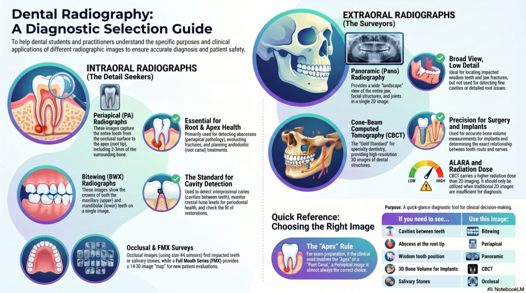
1. Intraoral Radiographs (The Detail Seekers)
🦷 Periapical (PA)
Primary Purpose: Shows the entire tooth from the occlusal surface to the apex (root tip) plus 2-3mm of surrounding bone.
- Detecting abscesses (periapical pathology).
- Evaluating root health and fractures.
- Endodontic (Root Canal) planning.
🥪 Bitewing (BWX)
Primary Purpose: Shows the crowns of both upper and lower teeth on one image.
- Detecting interproximal caries (cavities between teeth).
- Monitoring crestal bone levels (Periodontal health).
- Checking the fit of crowns or fillings.
Full Mouth Series (FMX)
A comprehensive “map” typically consisting of 14-20 images (PAs and BWs) for initial evaluation of a new patient.
Occlusal
Used to find impacted teeth, foreign bodies, or salivary stones. Uses a size #4 sensor/film.
2. Extraoral Radiographs (The Surveyors)
🎥 Panoramic (Pano)
A wide-view “landscape” of the entire jaw and facial structures.
- Locating impacted wisdom teeth.
- Evaluating growth and development.
- Detecting large lesions or jaw fractures.
Note: Panoramics are NOT used for detecting fine cavities or root details.
⚡ CBCT (Cone Beam 3D)
The “Gold Standard” for modern specialty dentistry.
- Implant Placement: Accurate measurements of bone volume.
- Oral Surgery: Seeing the exact relationship between roots and nerves.
Quick Summary Table
| If you need to see… | Use this Image: |
|---|---|
| Cavities between teeth | Bitewing |
| Abscess at the root tip | Periapical |
| Wisdom tooth position | Panoramic |
| 3D Bone Volume for Implants | CBCT |