Course Content
Introduction
Here is what to expect
0/3
Unit 1A: Purpose and Technique (50%) (Purpose)
A. Purpose of radiographic images 1. Periapical 2. Bitewing 3. Full mouth series 4. Occlusal 5. Full-mouth survey 6. Panoramic 7. Cephalometric 8. CBCT (cone-beam computed tomography) B. Technique 1. Review patient medical and dental histories for contraindications, including medications. 2. Intraoral techniques, including error correction. a. Paralleling b. Bisecting angle 3. Extraoral techniques, including error correction. a. Panoramic b. Cephalometric c. CBCT (cone-beam computed tomography) basics 4. Technique modifications based on anatomical variations and clinical conditions. 5. Purpose and maintenance of radiographic equipment. 6. Patient management techniques. 7. Mounting and anatomical landmarks that aid in mounting. 8. Anatomical structures and dental materials observed on images (e.g., differentiating between radiolucent and radiopaque areas). 9. Features of a diagnostically acceptable image. 10. Prepare images for legal requirements (e.g., HIPAA).
0/19
Unit 2A: Radiation production (25%)
Radiation production. 1. Sources of radiation for operators/other staff during radiation production. 2. Factors affecting x-ray production (e.g., kVp, mA, exposure time). 3. Radiation characteristics. 4. Radiation physics. a. Primary. b. Scatter (secondary). 5. Radiation biology. a. Short-and long-term effects of radiation on cells and tissues. b. Concepts of radiation dose.
0/8
Unit 2B: Radiation Safety
Radiation safety. 1. Causes of unnecessary exposure to radiation. 2. Patient exposure to radiation (ALARA, ADA recommendations). 3. Factors that influence radiation safety (e.g., filtration, shielding, collimation, PID length). 4. Patient radiation concerns. 5. Informed consent or patient refusal for exposure to radiation. 6. Protocol for suspected x-ray machine malfunctions.
0/9
Unit 3A: Standard precautions for equipment and supplies (25%)
Standard precautions for equipment and supplies according to ADA, CDC and OSHA, including but not limited to: 1. breakdown and setup of treatment room. 2. barriers. 3. position indicating and beam alignment devices. 4. clinical contact surfaces. 5. critical and semi-critical instrument sterilization
0/7
Unit 3B: Standard precautions for patients and operators
Standard precautions for patients and operators according to ADA, CDC and OSHA, including but not limited to: 1. hand hygiene. 2. PPE (donning, doffing). 3. cross contamination.
0/8
Vocabulary Terms
0/1
Practice Exams
0/1
RHS Exam Prep

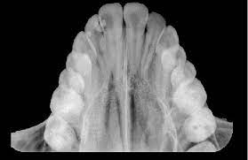
Occlusal Radiographs

The “Large Scale” Intraoral View

🎯 Learning Objectives

  • Identify the primary clinical indications for an occlusal radiograph.
  • State the specific film/sensor size used for adult occlusal imaging.
  • Describe the patient positioning and “sandwich” placement technique.
  • Differentiate between Maxillary and Mandibular occlusal perspectives.

1. Purpose: When to Use

An occlusal radiograph is used when a larger area of the arch needs to be visualized than a standard Periapical (PA) can provide. It is called “occlusal” because the patient “occludes” (bites down) on the sensor like a sandwich.

Impacted Teeth

Locating supernumerary (extra) teeth or impacted canines/wisdom teeth.

Pathology

Identifying large cysts, tumors, or fractures of the jawbone.

Salivary Stones

Locating “Sialoliths” (stones) in the floor of the mouth (Mandibular view).

2. Technique & Equipment

Feature Requirement
Film/Sensor Size Size 4 for adults (Size 2 for small children).
Patient Position Patient sits upright; the occlusal plane is parallel to the floor.
Vertical Angle Typically +65 degrees for Maxillary; -55 degrees for Mandibular.

🚨 DANB EXAM ALERT: Pediatric Use

Question: Why might a dentist choose an occlusal film over a Periapical for a 4-year-old?

Answer: Small children often have a shallow palate or sensitive gag reflex that prevents them from holding a standard PA sensor. The occlusal “sandwich” technique is much easier for young children to tolerate.

Exercise Files
104 Occlusal.mp3.mp3
Size: 0.00 B