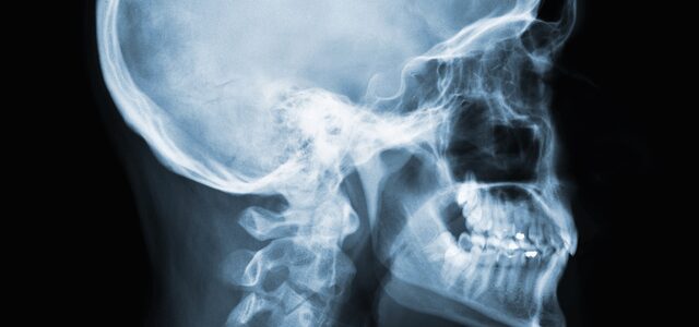
Cephalometric Radiography
Extraoral Imaging: The Lateral View
🎯 Learning Objectives
- Identify the primary clinical uses for cephalometric imaging.
- Describe the patient’s position relative to the sensor and X-ray beam.
- Identify the Cephalostat and its function.
- Recognize the anatomical landmarks visible on a lateral Ceph.
1. Purpose: The Orthodontic Map
A Cephalometric image is a standardized radiograph of the lateral (side) view of the skull. Unlike a Panoramic, which “unwraps” the jaw, a Ceph shows the relationship among the teeth, the jaw, and the soft-tissue profile.
Key Clinical Uses:
- Orthodontic Planning: Measuring the growth and development of the face.
- Surgical Evaluation: Planning for orthognathic (jaw) surgery.
- Soft Tissue Analysis: Viewing the patient’s profile to see how lip position relates to the teeth.
2. Technique & Equipment
To ensure orthodontic measurements are accurate, the patient’s head must be perfectly still and positioned at a specific distance from the X-ray source.
| Feature | Requirement |
|---|---|
| Orientation | The patient’s left side is usually placed against the sensor (Lateral view). |
| Cephalostat | A device with “ear rods” used to stabilize the head in a fixed position. |
| Midsagittal Plane | Must be perpendicular to the floor. |
🎓 DANB EXAM FOCUS: Identification
Question: Which extraoral image is best for evaluating a patient’s skeletal profile?
Answer: Cephalometric. If the question mentions “skeletal relationship,” “profile,” or “orthodontic measurements,” Cephalometric is the answer.