Bitewing Radiographs (BWX)
Domain: Purpose and Technique
🎯 Learning Objectives
- Identify the primary clinical purposes of the bitewing radiograph.
- List the specific anatomical structures that must be visible for a diagnostic BWX.
- Differentiate between horizontal and vertical bitewing applications.
- Identify and correct common bitewing technique errors (overlapping).
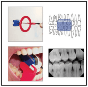
X-Ray holder placement for Bitewing is RED
1. Purpose of the Bitewing
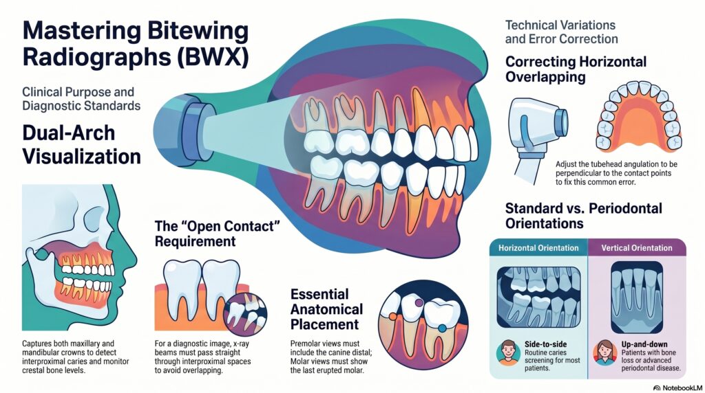
The bitewing radiograph is unique because it captures the crowns of both the maxillary and mandibular teeth on a single image. Its primary functions include:
Interproximal Caries
Detecting decay between teeth that cannot be seen clinically during a visual exam.
Crestal Bone Levels
Monitoring the height of the alveolar bone to screen for Periodontal Disease.
2. Diagnostic Criteria
For a bitewing to be considered “diagnostic,” it must meet the following visual standards:
- Open Contacts: The interproximal spaces must not be “overlapped.” The beam must pass straight through the contacts.
- Equal Coverage: An equal amount of Maxillary and Mandibular bone/crowns should be visible.
- Specific Placement:
- Premolar BWX: Must include the distal half of the Canine.
- Molar BWX: Must include the distal of the very last erupted molar.
3. Vertical vs. Horizontal Bitewings
While horizontal bitewings are standard, vertical bitewings are an essential modification for specific patients.
| Type | Orientation | Clinical Indication |
|---|---|---|
| Horizontal | Longer side placed side-to-side. | Routine caries screening for most patients. |
| Vertical | Longer side placed up-and-down. | Patients with bone loss or advanced periodontal disease. |
🚨 DANB EXAM ALERT: Overlapping
The most common error on a bitewing is Horizontal Overlapping.
The Cause: The central ray was not directed through the interproximal spaces.
The Fix: Re-adjust the tubehead’s horizontal angulation so it is perpendicular to the contact points.