Exercise Files
The “Large Scale” Intraoral View
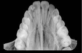
An occlusal radiograph is used when a larger area of the arch needs to be visualized than a standard Periapical (PA) can provide. It is called “occlusal” because the patient “occludes” (bites down) on the sensor like a sandwich.
Locating supernumerary (extra) teeth or impacted canines/wisdom teeth.
Identifying large cysts, tumors, or fractures of the jawbone.
Locating “Sialoliths” (stones) in the floor of the mouth (Mandibular view).
| Feature | Requirement |
|---|---|
| Film/Sensor Size | Size 4 for adults (Size 2 for small children). |
| Patient Position | Patient sits upright; the occlusal plane is parallel to the floor. |
| Vertical Angle | Typically +65 degrees for Maxillary; -55 degrees for Mandibular. |
Question: Why might a dentist choose an occlusal film over a Periapical for a 4-year-old?
Answer: Small children often have a shallow palate or sensitive gag reflex that prevents them from holding a standard PA sensor. The occlusal “sandwich” technique is much easier for young children to tolerate.