The Full Mouth Survey (FMX)
Module: Comprehensive Intraoral Imaging
🎯 Learning Objectives
- Define the standard film requirements for a complete adult survey.
- Apply the principles of Labial Mounting to organize images.
- Identify anatomical landmarks used to distinguish Maxillary vs. Mandibular views.
- Explain the clinical justification for an FMX under the ALARA principle.
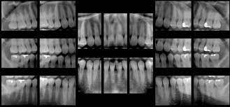
1. FMX Composition
A standard adult FMX typically consists of 18 to 20 images. This ensures that every tooth, every contact point, and every root apex is captured from at least one angle.
| Image Type | Quantity | Target Area |
|---|---|---|
| Anterior PAs | 6 – 8 | Centrals, Laterals, and Canines. |
| Posterior PAs | 8 | Premolars and Molars (all 4 quadrants). |
| Bitewings | 4 | Interproximal decay screening. |
2. Mounting: The Labial Method
The DANB exam focuses on Labial Mounting. In this method, radiographs are placed in the mount as if you are standing outside the mouth looking in.
- The “Dot”: The embossed identification dot on the film/sensor must be raised (convex) toward the viewer.
- Perspective: The patient’s left is your right; the patient’s right is your left.
- Anatomy: Maxillary teeth are always mounted with roots pointing up; Mandibular teeth with roots pointing down.
3. The Curve of Spee
When mounting posterior bitewings, look for the Curve of Spee. This is the natural upward curve of the occlusal plane as it moves toward the back of the mouth.
- If the curve looks like a smile, the images are mounted correctly.
- If the curve looks like a frown, the images are upside down or swapped.
🎓 DANB EXAM FOCUS: Edentulous Surveys
You may be asked if an FMX is necessary for a patient with no teeth. Yes. For an edentulous patient, the number of images is typically reduced (usually to 14 PAs), but the survey is required to detect retained roots, cysts, or systemic bone changes.