Course Content
Introduction
Here is what to expect
0/3
Unit 1A: Purpose and Technique (50%) (Purpose)
A. Purpose of radiographic images 1. Periapical 2. Bitewing 3. Full mouth series 4. Occlusal 5. Full-mouth survey 6. Panoramic 7. Cephalometric 8. CBCT (cone-beam computed tomography) B. Technique 1. Review patient medical and dental histories for contraindications, including medications. 2. Intraoral techniques, including error correction. a. Paralleling b. Bisecting angle 3. Extraoral techniques, including error correction. a. Panoramic b. Cephalometric c. CBCT (cone-beam computed tomography) basics 4. Technique modifications based on anatomical variations and clinical conditions. 5. Purpose and maintenance of radiographic equipment. 6. Patient management techniques. 7. Mounting and anatomical landmarks that aid in mounting. 8. Anatomical structures and dental materials observed on images (e.g., differentiating between radiolucent and radiopaque areas). 9. Features of a diagnostically acceptable image. 10. Prepare images for legal requirements (e.g., HIPAA).
0/19
Unit 2A: Radiation production (25%)
Radiation production. 1. Sources of radiation for operators/other staff during radiation production. 2. Factors affecting x-ray production (e.g., kVp, mA, exposure time). 3. Radiation characteristics. 4. Radiation physics. a. Primary. b. Scatter (secondary). 5. Radiation biology. a. Short-and long-term effects of radiation on cells and tissues. b. Concepts of radiation dose.
0/8
Unit 2B: Radiation Safety
Radiation safety. 1. Causes of unnecessary exposure to radiation. 2. Patient exposure to radiation (ALARA, ADA recommendations). 3. Factors that influence radiation safety (e.g., filtration, shielding, collimation, PID length). 4. Patient radiation concerns. 5. Informed consent or patient refusal for exposure to radiation. 6. Protocol for suspected x-ray machine malfunctions.
0/9
Unit 3A: Standard precautions for equipment and supplies (25%)
Standard precautions for equipment and supplies according to ADA, CDC and OSHA, including but not limited to: 1. breakdown and setup of treatment room. 2. barriers. 3. position indicating and beam alignment devices. 4. clinical contact surfaces. 5. critical and semi-critical instrument sterilization
0/7
Unit 3B: Standard precautions for patients and operators
Standard precautions for patients and operators according to ADA, CDC and OSHA, including but not limited to: 1. hand hygiene. 2. PPE (donning, doffing). 3. cross contamination.
0/8
Vocabulary Terms
0/1
Practice Exams
0/1
RHS Exam Prep

Cephalometric Radiography

Extraoral Imaging: The Lateral View

🎯 Learning Objectives

  • Identify the primary clinical uses for cephalometric imaging.
  • Describe the patient’s position relative to the sensor and X-ray beam.
  • Identify the Cephalostat and its function.
  • Recognize the anatomical landmarks visible on a lateral Ceph.

1. Purpose: The Orthodontic Map

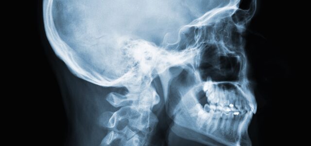
A Cephalometric image is a standardized radiograph of the lateral (side) view of the skull. Unlike a Panoramic, which “unwraps” the jaw, a Ceph shows the relationship among the teeth, the jaw, and the soft-tissue profile.

Key Clinical Uses:

  • Orthodontic Planning: Measuring the growth and development of the face.
  • Surgical Evaluation: Planning for orthognathic (jaw) surgery.
  • Soft Tissue Analysis: Viewing the patient’s profile to see how lip position relates to the teeth.

2. Technique & Equipment

To ensure orthodontic measurements are accurate, the patient’s head must be perfectly still and positioned at a specific distance from the X-ray source.

Feature Requirement
Orientation The patient’s left side is usually placed against the sensor (Lateral view).
Cephalostat A device with “ear rods” used to stabilize the head in a fixed position.
Midsagittal Plane Must be perpendicular to the floor.

🎓 DANB EXAM FOCUS: Identification

Question: Which extraoral image is best for evaluating a patient’s skeletal profile?

Answer: Cephalometric. If the question mentions “skeletal relationship,” “profile,” or “orthodontic measurements,” Cephalometric is the answer.

Exercise Files
107 Cephalometric.mp3.mp3
Size: 0.00 B