Course Content
Introduction
Here is what to expect
0/3
Unit 1A: Purpose and Technique (50%) (Purpose)
A. Purpose of radiographic images 1. Periapical 2. Bitewing 3. Full mouth series 4. Occlusal 5. Full-mouth survey 6. Panoramic 7. Cephalometric 8. CBCT (cone-beam computed tomography) B. Technique 1. Review patient medical and dental histories for contraindications, including medications. 2. Intraoral techniques, including error correction. a. Paralleling b. Bisecting angle 3. Extraoral techniques, including error correction. a. Panoramic b. Cephalometric c. CBCT (cone-beam computed tomography) basics 4. Technique modifications based on anatomical variations and clinical conditions. 5. Purpose and maintenance of radiographic equipment. 6. Patient management techniques. 7. Mounting and anatomical landmarks that aid in mounting. 8. Anatomical structures and dental materials observed on images (e.g., differentiating between radiolucent and radiopaque areas). 9. Features of a diagnostically acceptable image. 10. Prepare images for legal requirements (e.g., HIPAA).
0/19
Unit 2A: Radiation production (25%)
Radiation production. 1. Sources of radiation for operators/other staff during radiation production. 2. Factors affecting x-ray production (e.g., kVp, mA, exposure time). 3. Radiation characteristics. 4. Radiation physics. a. Primary. b. Scatter (secondary). 5. Radiation biology. a. Short-and long-term effects of radiation on cells and tissues. b. Concepts of radiation dose.
0/8
Unit 2B: Radiation Safety
Radiation safety. 1. Causes of unnecessary exposure to radiation. 2. Patient exposure to radiation (ALARA, ADA recommendations). 3. Factors that influence radiation safety (e.g., filtration, shielding, collimation, PID length). 4. Patient radiation concerns. 5. Informed consent or patient refusal for exposure to radiation. 6. Protocol for suspected x-ray machine malfunctions.
0/9
Unit 3A: Standard precautions for equipment and supplies (25%)
Standard precautions for equipment and supplies according to ADA, CDC and OSHA, including but not limited to: 1. breakdown and setup of treatment room. 2. barriers. 3. position indicating and beam alignment devices. 4. clinical contact surfaces. 5. critical and semi-critical instrument sterilization
0/7
Unit 3B: Standard precautions for patients and operators
Standard precautions for patients and operators according to ADA, CDC and OSHA, including but not limited to: 1. hand hygiene. 2. PPE (donning, doffing). 3. cross contamination.
0/8
Vocabulary Terms
0/1
Practice Exams
0/1
RHS Exam Prep

Cone Beam Computed Tomography (CBCT)

The Transition to 3D Imaging

🎯 Learning Objectives

  • Define CBCT and how it differs from traditional 2D radiography.
  • Identify the clinical indications for a 3D scan (Implants, Endodontics).
  • Understand the concept of Voxels vs. Pixels.
  • Compare radiation doses between CBCT and standard FMX.
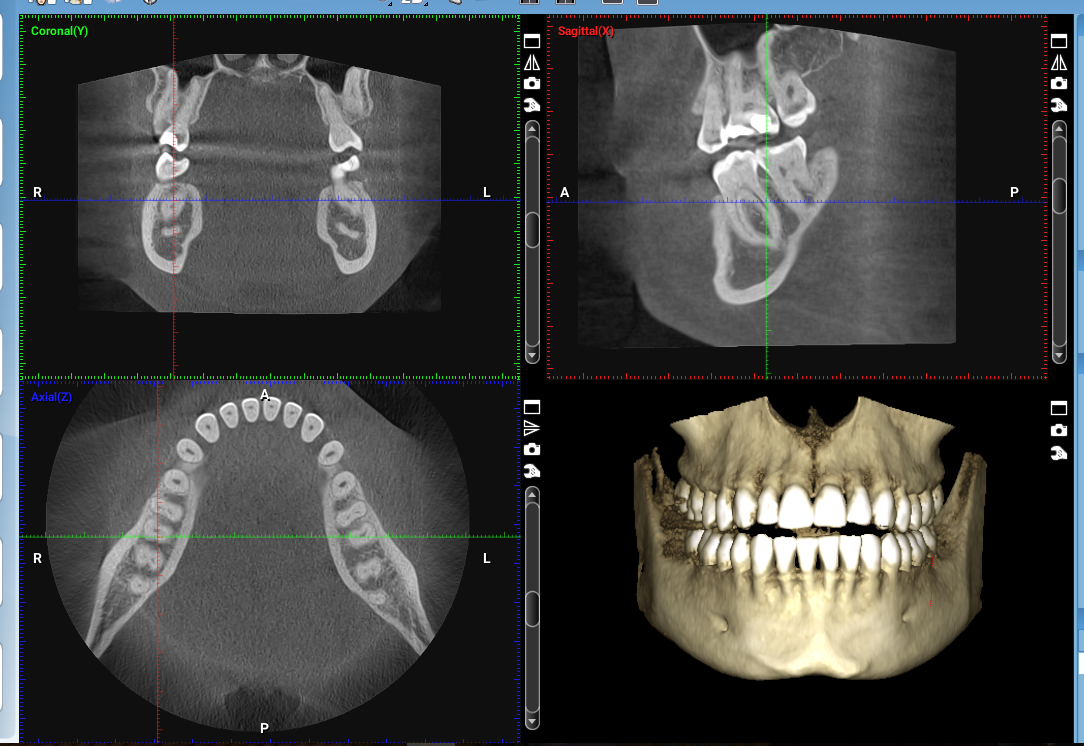
1. What is CBCT?

Unlike a traditional X-ray that provides a flat, two-dimensional (2D) view where structures overlap, CBCT uses a cone-shaped X-ray beam to capture a series of images that a computer reconstructs into a three-dimensional (3D) model.

The Power of 3D:

CBCT allows the dentist to view the “depth” of anatomy. This is critical for seeing exactly where a nerve is located or measuring the width of bone before surgery.

2. Pixels vs. Voxels

The RHS exam loves technical definitions. You must know the difference between the building blocks of these images:

  • Pixel: A 2D digital unit (found on sensors and Pano images).
  • Voxel: A 3D digital unit (found in CBCT scans). Think of a Voxel as a pixel with volume.

3. Clinical Indications

Because CBCT carries a higher radiation dose than a single PA, it is only used when 2D imaging is insufficient:

Specialty Use Case
Implantology Measuring bone volume and density for implant placement.
Endodontics Locating extra canals or vertical root fractures.
Oral Surgery Mapping the Mandibular Nerve before wisdom tooth extraction.

🎓 DANB EXAM FOCUS: DICOM Images

The universal file format for CBCT and medical imaging is DICOM (Digital Imaging and Communications in Medicine). You may see this term in questions regarding how images are shared between specialists.

Exercise Files
108 CBCT.mp3.mp3
Size: 0.00 B