Course Content
Introduction
Here is what to expect
0/3
Unit 1A: Purpose and Technique (50%) (Purpose)
A. Purpose of radiographic images 1. Periapical 2. Bitewing 3. Full mouth series 4. Occlusal 5. Full-mouth survey 6. Panoramic 7. Cephalometric 8. CBCT (cone-beam computed tomography) B. Technique 1. Review patient medical and dental histories for contraindications, including medications. 2. Intraoral techniques, including error correction. a. Paralleling b. Bisecting angle 3. Extraoral techniques, including error correction. a. Panoramic b. Cephalometric c. CBCT (cone-beam computed tomography) basics 4. Technique modifications based on anatomical variations and clinical conditions. 5. Purpose and maintenance of radiographic equipment. 6. Patient management techniques. 7. Mounting and anatomical landmarks that aid in mounting. 8. Anatomical structures and dental materials observed on images (e.g., differentiating between radiolucent and radiopaque areas). 9. Features of a diagnostically acceptable image. 10. Prepare images for legal requirements (e.g., HIPAA).
0/19
Unit 2A: Radiation production (25%)
Radiation production. 1. Sources of radiation for operators/other staff during radiation production. 2. Factors affecting x-ray production (e.g., kVp, mA, exposure time). 3. Radiation characteristics. 4. Radiation physics. a. Primary. b. Scatter (secondary). 5. Radiation biology. a. Short-and long-term effects of radiation on cells and tissues. b. Concepts of radiation dose.
0/8
Unit 2B: Radiation Safety
Radiation safety. 1. Causes of unnecessary exposure to radiation. 2. Patient exposure to radiation (ALARA, ADA recommendations). 3. Factors that influence radiation safety (e.g., filtration, shielding, collimation, PID length). 4. Patient radiation concerns. 5. Informed consent or patient refusal for exposure to radiation. 6. Protocol for suspected x-ray machine malfunctions.
0/9
Unit 3A: Standard precautions for equipment and supplies (25%)
Standard precautions for equipment and supplies according to ADA, CDC and OSHA, including but not limited to: 1. breakdown and setup of treatment room. 2. barriers. 3. position indicating and beam alignment devices. 4. clinical contact surfaces. 5. critical and semi-critical instrument sterilization
0/7
Unit 3B: Standard precautions for patients and operators
Standard precautions for patients and operators according to ADA, CDC and OSHA, including but not limited to: 1. hand hygiene. 2. PPE (donning, doffing). 3. cross contamination.
0/8
Vocabulary Terms
0/1
Practice Exams
0/1
RHS Exam Prep

The DANB RHS Exam Overview

The Radiation Health and Safety (RHS) exam is a national requirement for dental assistants seeking to advance their careers. Whether you are aiming for your CDA or just need your state’s X-ray certification, this is the hurdle you must clear. 
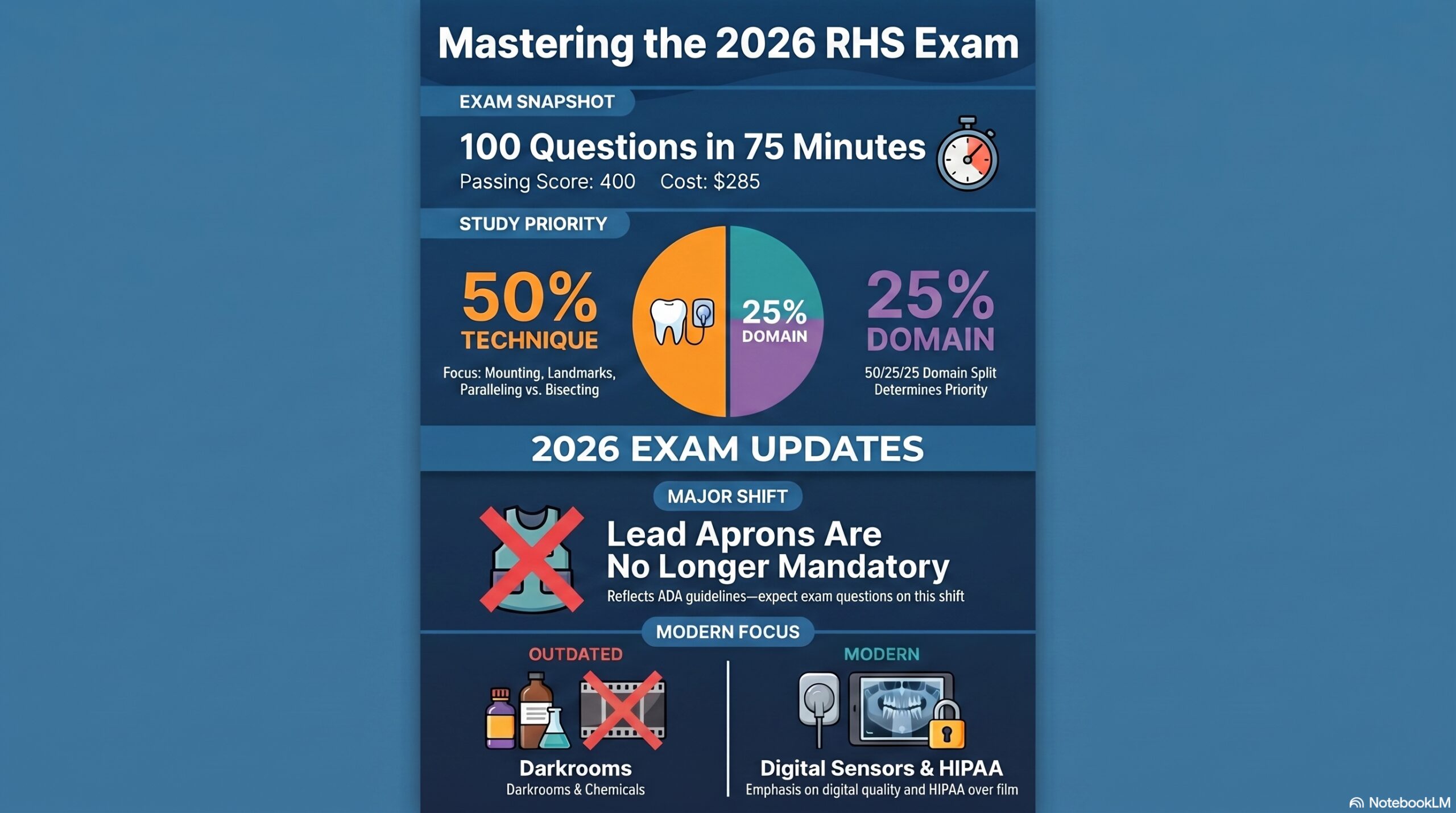
Exam Feature Details (2026 Standards)
Number of Questions 100 Multiple Choice
Time Allotted 75 Minutes
Passing Score 400 (on a 200-900 scale)
Standard Fee $285
⚠️ 2026 EXAM UPDATE:
The DANB RHS exam now reflects the ADA/AAOMR guidelines which state that lead aprons and thyroid collars are no longer mandatory for routine dental radiographs. Expect questions regarding this change in the Safety domain!

Exam Domain Breakdown

The exam is split into three weighted categories. Your study time should mirror these percentages:

50% Technique

Mounting, Error Correction, Landmarks, and Paralleling vs. Bisecting.

25% Safety

ALARA principle, biological effects, and operator protection.

25% Quality

Infection control (Digital sensors) and Legal/HIPAA requirements.

Key Success Tips

  • Think Digital: Darkrooms and chemicals are largely gone from the exam. Focus on sensors.
  • Learn the Landmarks: You will be shown digital images and asked to identify the Maxillary Sinus, Mental Foramen, etc.
  • Practice the Clock: You have less than 1 minute per question. Use our mock exams to build speed.